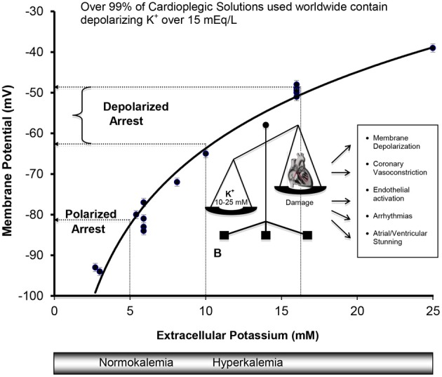
Figure 3.

Cell Membranes of the Atria, Purkinje, and Ventricular Myocytes in vivo behave as a K+ electrode over a range of extracellular K+ from 3 to 25 mM. Data were obtained from isolated rat, rabbit, and guinea-pig hearts (Kleber, 1983; Masuda et al., 1990; Sloots and Dobson, 2010; Dobson and Jones, 2004; Dobson, 2010) and from isolated cells (Wan et al., 2000). The membrane potential φ (mV) = 27 ln [K+] − 126, R2 = 0.97, where [K+] is extracellular potassium concentration in mM. Membrane potentials (φ) were measured directly using potassium microelectrodes (Kleber, 1983; Masuda et al., 1990; Snabaitis et al., 1997; Wan et al., 2000) or calculated from Nerstian distributions of potassium (Dobson and Jones, 2004; Sloots and Dobson, 2010). The relation between potassium and membrane potential for ventricular muscle also agree well with microelectrode measurements on isolated human atrial muscle (Gelband et al., 1972), and for isolated purkinje fibers above 5.4 mM K+ bathed in Tyrode's solution (Sheu et al., 1980).
