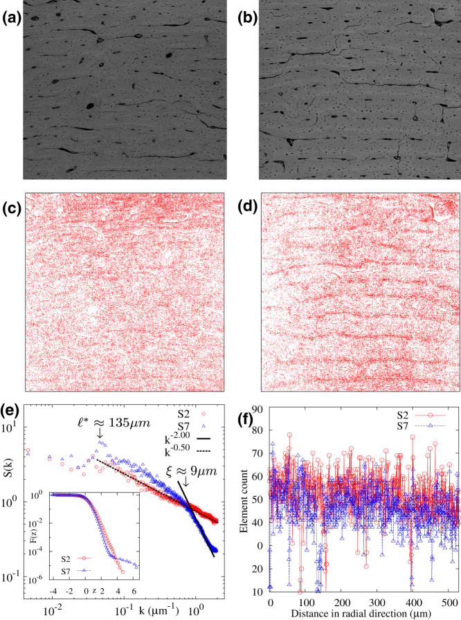
Figure 3. BSE images of sections (a) S2 anterior and (b) S7 anterior, where the field width is 1475 μm (966 pixels).
The brightest 10% pixels of the BSE images for (c) S2 anterior and (d) S7 anterior. (e) Structure factor S(k) along the direction perpendicular to the banding. Inset: F(z), the probability that the scaled gray scale value is greater than z for S2 and S7. (f) EDS data for calcium content along a line perpendicular to the banding.

