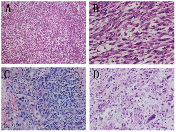
Figure 3. Pathological findings of 3 MFH subtypes.

Storiform-pleomorphic subtype: (A) The tumor was composed of hypercellular spindle cells arranged in a characteristic storiform pattern (H&E 100×). (B) Amplification of Figure 3A, shows atypical, highly proliferating cells (H&E 400×). Inflammatory subtype: (C) Proliferation of fibroblasts and multinucleated histiocytes. Numbers of inflammatory cells were seen in the interstitial substance (H&E 400×). Giant cell subtype: (D) Numbers of spindle-shaped or tadpole-shaped, multinucleated giant cells were seen. Cell nuclei ranged from 2 to 14, and the shape was irregular (H&E 400×).
