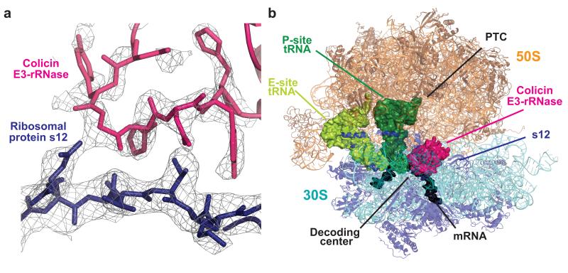
Figure 1. Structure of colicin E3-rRNase bound to the 70S ribosome.
(a) Representative electron density from a 3mFo-2dFc map displayed at 0.9 σ, with the refined model of E3-rRNase (pink) and ribosomal protein S12 (blue). (b) Overall view of the complex, with colicin E3-rRNase and P-site and E-site tRNA depicted as surfaces, and rRNA and proteins as cartoons.

