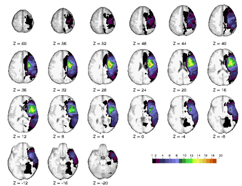
Fig 1.

Horizontal slices of anatomical MRI standardized in Talairach atlas showing the lesion distribution for 30 representative patients, obtained at the chronic testing session. The color scale represents the number of patients with damage in a specific voxel.
