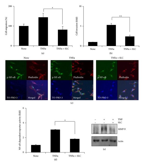
Figure 1.

SLC inhibits TNFα-induced MDA-MB-231 cell migration and invasion by inhibiting NF-κB activation. (a) Cell migration was measured by wound healing assay. MDA-MB-231 cells were seeded and scratched, pretreated with SLC for 1 hour, and then exposed to TNFα for 24 hours. Cell migration was determined by counting cell numbers migrated from the wound healing region. *P < 0.05. (b) MDA-MB-231 cells were seeded on the upper chambers and pretreated with SLC for 1 hour and then exposed to TNFα for 24 hours. Invading cells were stained with hematoxylin and eosin, and the cell numbers were measured. **P < 0.001. (c) MDA-MB-231 cells were pretreated with SLC for 1 hour, then exposed to TNFα for 6 hours, and stained with p-NF-κB antibody. Phalloidin and TO-PRO-3 were for staining F-actin and the nucleus, respectively. The object was 20x, and scale bars on the image indicate 50 μm. (d) MDA-MB-231 cells were transfected with the NF-κB-dependent luciferase reporter, pretreated with SLC for 1 hour, and then exposed to TNFα for 6 hours. Luciferase assays were done by using dual-luciferase reporter assay. All transfections included the RLTK-Luc for transfection efficiency. *P < 0.05. (e) MDA-MB-231 cells were pretreated with SLC for 1 hour and then exposed to TNFα for 24 hours. MMP-9 protein was measured by western blotting. Tubulin was used for the loading control.
