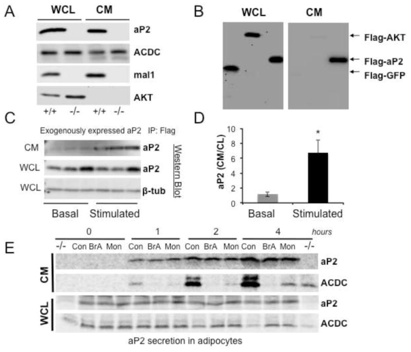
Figure 1. Secretion of aP2 in vitro from cultured cells.

a, aP2 secretion in adipocytes. Whole cell lysate (WCL) and conditioned medium (CM) from differentiated WT (+/+) or AM−/− (−/−) adipocytes lacking both aP2 and mal1 were blotted using anti-aP2, adiponectin (ACDC), mal1, or AKT antibodies. These cell lines were developed in-house. b, aP2 secretion in HEK 293 cells. Whole cell lysates (WCL) or immunoprecipated conditioned medium (CM) from HEK 293 cells transfected with Flag-AKT, Flag-GFP-aP2 or Flag-GFP plasmids were used to detect aP2 by western blotting with an anti-Flag antibody. c, CMV promoter driven aP2 expression and its regulated secretion studied in aP2−/− adipocytes. Flag-aP2 was transfected into aP2−/− cells and its appearance in the CM was examined under basal and forskolin (FSK) stimulated condition (20μM for 1h). CM samples were immunoprecipitated with an anti-Flag antibody and blotted with anti-aP2. Cell lysate was probed with aP2 and β-tubulin (β-tub) antibodies. d, Quantitation of data shown in panel c determined by using ImageJ program. * p < 0.05 in student’s t test. e, Pulse chase analysis of aP2 secretion. Cultured adipocytes were metabolically labeled and then treated with vehicle, brefeldin A (BrA, 10 μg/ml) or monensin (Mon, 5 μM), and samples were taken at indicated time points. Proteins in CM were immunoprecipitated using anti-aP2 or adiponectin (ACDC) antibodies. After electrophoresis, radiolabeled proteins were subjected to autoradiography. An aP2−/− cell lysate was used as negative control. Data are presented as means ± SEM.
