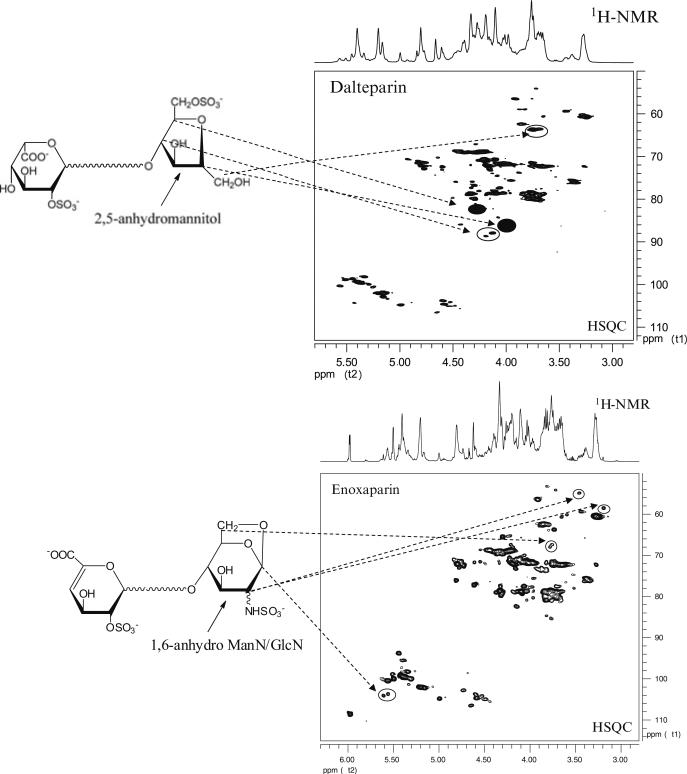
Fig. 4.
HSQC spectra of low-molecular-weight heparins. 2D-NMR spectra of dalteparin and enoxaparin exhibit unique cross peaks arising from signature structures. These structures result from the specific chemistry used to depolymerize unfractionated heparin into lower molecular weight chains. The 2,5 anhydromannitol in dalteparin, and the 1,6-anhydro structure in enoxaparin, along with their associated signature cross peaks are shown

