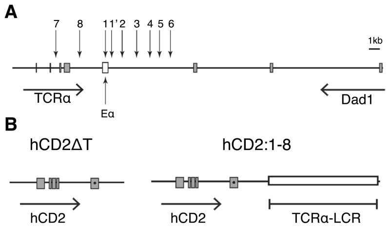
Figure 1. The TCRα LCR genomic region and reporter transgenes.
(A) Diagram of the TCRα/Dad1 locus. Vertical arrows depict DNase hypersensitive sites (HS)1-8 of the TCRα LCR. The open box marks the Eα classical transcriptional enhancer. All other boxes indicate exons of their respective genes. Horizontal arrows indicate the transcription orientation of the genes. Diagram is drawn to scale. (B) Depiction of the hCD2ΔT and hCD2:1-8 transgenes. A premature stop codon (●) was introduced in exon V prior to the codons of the cytoplasmic tail (24) In hCD2:1-8, the TCRα LCR cassette (18) (open box) containing an exon-free HS1-8 fragment is linked to hCD2ΔT gene fragment.

