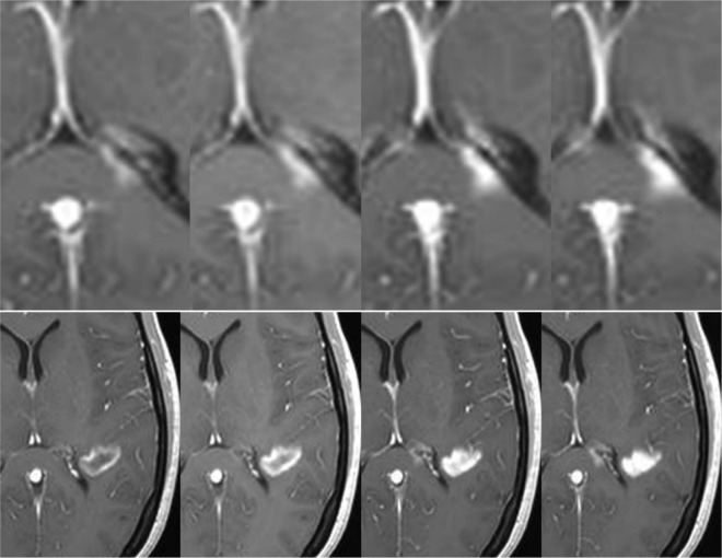
Figure 2.
Evolution of contrast uptake in a newly formed lesion. Serial contrast-enhanced T1-weighted images obtained 5, 10, 15, and 20 min after gadolinium injection. A nodular-enhanced lesion located in the splenium of the corpus callosum (upper row) increases in size over time (centrifugal pattern), while an initial ring-enhanced lesion (lower row) becomes nodular (centripetal pattern).

