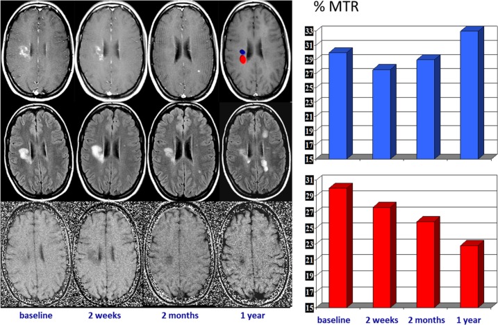
Figure 6.
Serial magnetic resonance imaging changes occurring in an acute multiple sclerosis plaque located in the right centrum ovale. Contrast-enhanced T1-weighted images (upper row), T2-FLAIR images (middle row), and magnetization transfer (MTR) maps (lower row) obtained at baseline and at 7 days, 1 month, and 1 year later. The acute enhanced plaque shows the typical waxing and waning characteristics on T2-weighted images. The initial T2 lesion shrinkage observed after cessation of contrast uptake can be interpreted as a consequence of resorption of inflammatory edema, but the subsequent extended size decrease likely reflects a repair process. The serial MTR maps show two different components within the lesion, one with partial MTR recovery, likely reflecting remyelination, and the other with a progressive MTR decrease, likely reflecting ongoing demyelination (on the right, a plot of the serial MTR values obtained at the two locations).
FLAIR, fluid attenuation inversion recovery.

