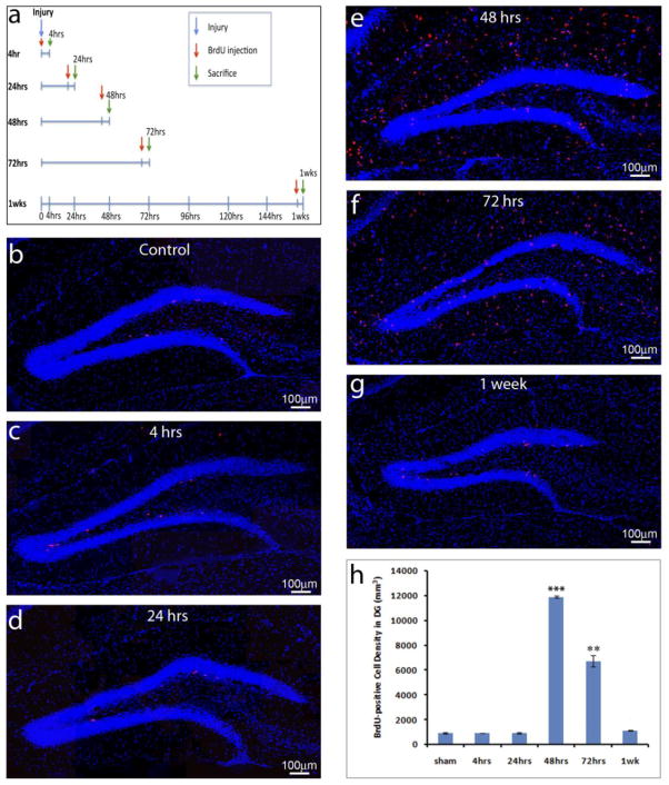
Figure 1. TBI insults promote NSC proliferation in the adult hippocampus.
(a) The schematic shows pulse labeling of proliferating cells in the hippocampus at different days following traumatic brain injury (TBI). (b–g) Immunostaining with an anti- BrdU antibody (red) to identify proliferating cells in the hippocampus of sham control (b) or TBI mice at different time points after surgery (c–g). Nuclei are stained with DAPI (blue) to show the structure of the dental gyrus. (h) Quantification of BrdU-positive cells in the hippocampal dentate gyrus following TBI. (*** p<0.0001; ** p<0.001, n=5 for each group).

