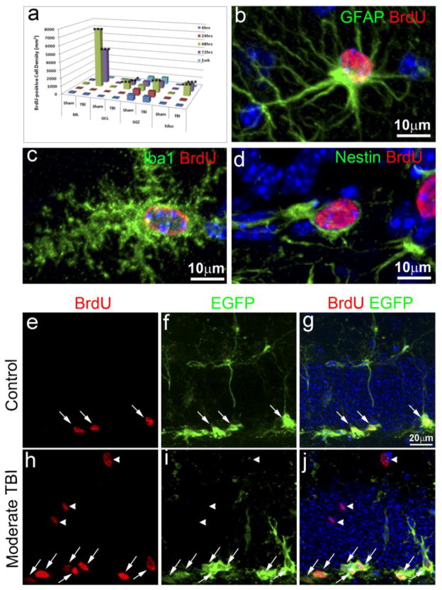
Figure 2. Spatial and temporal distribution of the proliferating cells and their fate in the hippocampus one week after TBI.
(a) Quantification of the proliferating cells in the hippocampus after traumatic brain injury (TBI) and their spatial and temporal distribution (n=5). * p<0.05; ** p<0.001; *** p<0.0001. (b) Double immunostaining with anti-BrdU (red) and anti-GFAP (green) antibodies to identify proliferating astrocytes in the injured hippocampus. (c) Double immunostaining with anti-BrdU (red) and Iba1 (green) antibodies to identify proliferating microglia in the injured hippocampus. (d) Double immunostaining with anti-BrdU (red) and anti-nestin (green) antibodies to identify proliferating neural stem/progenitor cells (NSCs) in the injured hippocampus. (e) Immunostaining with anti-BrdU (red) to identify proliferating cells in the hippocampal dentate gyrus (HDG) of nestin-EGFP transgenic mice after sham treatment. (f) Immunostaining with anti-EGFP (green) to identify EGFP expressing NSCs in the HDG of nestin-EGFP transgenic mice after sham treatment. (g). Merged image of (e) and (f). The BrdU and EGFP double-positive cells are pointed out by white arrows. (h) Immunostaining with anti-BrdU (red) to identify proliferating cells in the hippocampal dentate gyrus of nestin-EGFP transgenic mice after TBI. (i) Immunostaining with anti-EGFP (green) to identify EGFP expressing NSCs in the HDG of nestin-EGFP transgenic mice after TBI. (j). Merged image of (h) and (i). The BrdU and EGFP double-positive cells are pointed out by white arrows. The BrdU+ positive cells without expressing EGFP are pointed out by white arrowheads.

