
Figure 4. Astrocyte activation in the hippocampal dentate gyrus following TBI.
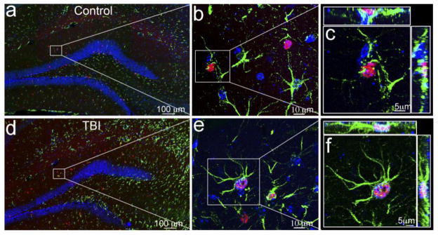
Double immunostaining with anti-BrdU (red) and anti-GFAP (green) antibodies to identify the proliferating astrocytes in the hippocampus of the sham (a–c) or traumatic brain injury (TBI) mice (d–f). The nuclei are stained with DAPI (blue) to show the hippocampal dentate gyrus (HDG) structure. (a, d) Proliferating astrocytes in the hippocampus of the sham or TBI mice at low magnification. BrdU-positive cells appear in red and GFAP-positive cells in green. (b, e) BrdU-positive astrocytes (indicated by the white box) in the molecular layer of sham or TBI mice at high magnification. (c, f) Fluorescent images with 3-dimensional reconstruction to confirm the proliferating astrocytes (indicated by a white box) in the molecular layer of sham or TBI mice.