Figure 6.
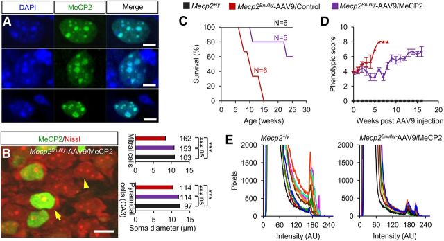
MeCP2 expressed from virus binds to DNA, restores normal neuronal somal size, and improves survival. A, Ectopic MeCP2 localizes to DAPI+ heterochromatin puncta in Mecp2Bnull/y-scAAV9/MeCP2–injected mice. Shown is colocalization of DAPI and MeCP2 in olfactory neuron (top), CA3 pyramidal neuron (middle), and dentate gyrus astrocyte (bottom). Pearson's correlation coefficient = 0.943, 0.932, and 0.985, respectively. Scale bar, 5 μm. B, Image (left) shows representative MeCP2-positive (arrow) and MeCP2-negative (arrowhead) CA3 pyramidal neurons. Scale bar, 10 μm. Average somal diameters (right) of MeCP2-positive (purple) and MeCP2-negative (red) CA3 pyramidal neurons and olfactory bulb mitral cells from Mecp2Bnull/y-scAAV9/MeCP2–injected mice (n = 2). Also shown are measurements from WT mice (black bars; n = 2). The number of cells analyzed is indicated above each bar. ***p < 0.001 by one-way ANOVA (Newman–Keuls multiple-comparison test). Data are means ± SEM. C, Kaplan-Meier survival curve. D, Observational scores. Mecp2Bnull/y-scAAV9/MeCP2 (n = 5), Mecp2Bnull/y-AAV9/Control (n = 6), and Mecp2+/y (n = 6). Data are means ± SEM. E, Field pixel intensities of MeCP2-Cy3 immunofluorescence measured from brainstem sections of Mecp2+/y and Mecp2Bnull/y-scAAV9/MeCP2 mice. Traces represent pixel intensities from individual fields and each field is indicated by a differently colored trace, n = 2 mice per genotype, 5 fields per mouse.

