Figure 2.
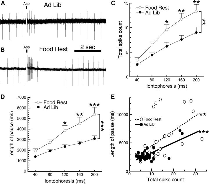
Food restriction increases the sensitivity of DA neurons to aspartic acid. Firing of substantia nigra DA neurons was monitored using loose cell-attached recordings. A, A sample trace shows that an 80 ms iontophoretic pulse of aspartic acid (indicated by a small black bar) in a neuron from an ad libitum-fed mouse produced a brief increase in firing rate followed by a pause. B, When the same pulse was applied to a DA neuron from a food-restricted mouse, it produced more action potentials followed by a longer pause when compared with ad libitum controls. C, D, DA neurons from food-restricted mice exhibited a greater number of spikes (C) and a longer pause length (D) across a range (40–200 ms) of iontophoretic applications of aspartic acid. E, Analysis of the physiological consequences of the 200 ms ejection indicated that total spike count and pause length were significantly correlated in cells from both AL and FR mice; however, the two slopes were not significantly different from each other. *p < 0.05, **p < 0.01, ***p < 0.001. Food Rest, food restricted; Ad Lib, ad libitum.

