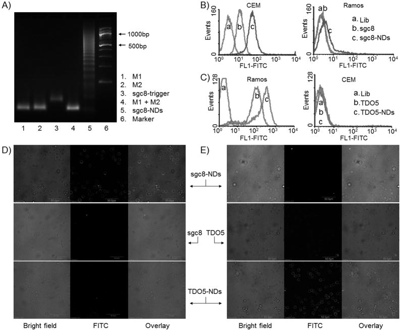
Figure 1.
Selective anchoring of chemically-modified fluorescent DNA nanodevices on target cell surfaces. A, An agarose gel electrophoresis image verifying the self-assembly of sgc8-NDs upon the initiation of sgc8-trigger. M1 and M2 (Lane 4) did not react unless sgc8-trigger was present. B–C, Flow cytometric results indicating that fluorescent sgc8-and TDO5-nanodevices were selectively anchored on the surfaces of CEM cells (B) and Ramos cells (C), respectively, but not the corresponding nontarget cells. Fluorescent nanodevice-anchored target cells displayed enhanced fluorescence intensities, compared to cells bound by the corresponding aptamers only. D–E, Confocal microscopy images indicating that fluorescent sgc8- and TDO5-NDs were selectively anchored on the surfaces of CEM cells (D) and Ramos cells (E), respectively. (Scale bar: 50 μm; Lib: random sequences; lib, sgc8, TDO5, M1, M2: labeled with FITC.)

