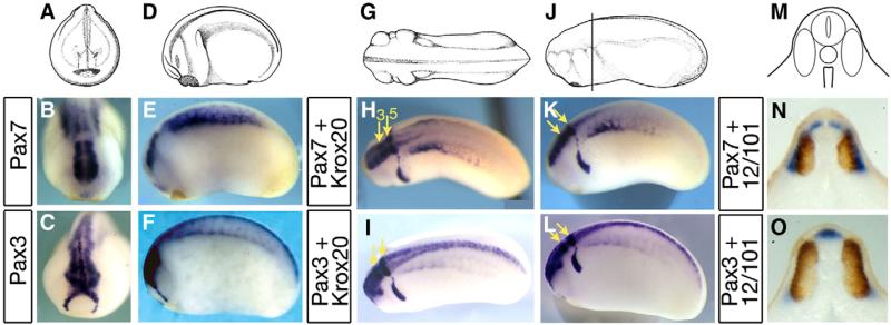
Fig. 2.
Distinct pax7 and pax3 patterns in central nervous system and paraxial mesoderm at tailbud stages. (A–C) Front views of stage 22 embryos (A) show pax7 expression in the caudal part of the brain (B), whereas pax3 labels both the whole brain and the hatching gland (C). (D–F) Side views (D) illustrate pax7 restriction to mesencephalon, hindbrain and anterior spinal cord whereas pax3 is found along the entire anterior–posterior length of the central nervous system. Pax7 labels the central paraxial mesoderm while pax3 is found in hypaxial cells. (G–L) Dorsal (G–I) and side (J–L) views of tailbud stage 24, with double staining for pax3 or pax7 and krox20, which labels hindbrain rhombomeres r3 and r5 (yellow arrows), confirms the limited extent of pax7 expression along the spinal cord. (M–O) Tailbud stage 24 transverse sections were double-stained with 12–101 myotome marker (brown). This shows pax7 expression in the alar plate of the anterior-most spinal cord and medial myotome (N) and pax3 in the roof plate, alar plates and in the hypaxial myotome (O).

