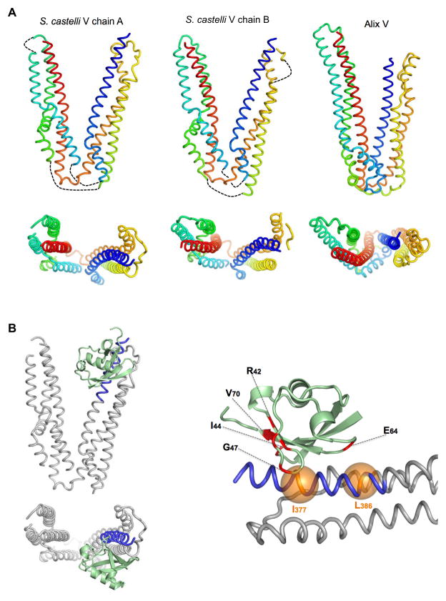
Figure 4. Crystal structure of the Bro1 V domain in complex with Ub.
A. 3.6Å structures of the Bro1 V domains found within the asymmetric unit (PDB ID: 4JIO). A cartoon model of the Alix V domain (PDB ID:2OEX) is shown at right. Below is alternate view from the top of the arms, looking into the vertex of the two Bro1 V domain structures together with Alix V. The backbone is colored blue-to-red according to amino acid order (N- to C-terminus). Loops not resolved in electron density maps are represented by dotted lines.
B. Structure of the Ub:Bro1 V domain complex, showing Ub (green) bound to the N-terminus of the Bro1 V domain within the first trihelical bundle (residues 370–392: blue). Model at right is closer view of Ub:V interaction. Ub residues that undergo significant NMR chemical perturbation upon Bro1 V binding are colored red. Positions of the Bro1 V I377 and L386 Cα atoms are shown in orange.

