
Figure 4.
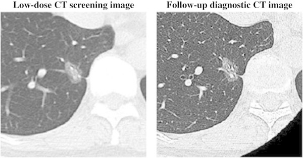
Low-dose CT screening image and follow-up diagnostic CT image. Low-dose CT screening image with a slice thickness and interval of 3 mm. Follow-up diagnostic CT image with an image slice thickness and interval of 1 mm. A 60-year-old woman with bronchioalveolar carcinoma (adenocarcinoma in situ) as mixed ground-glass opacity or pure ground-glass opacity in the right lower lobe (T1N0M0).