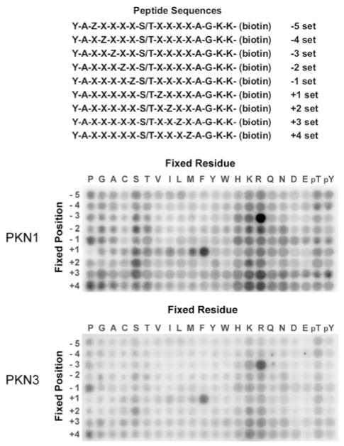
Figure 1. Determination of phosphorylation motifs for PKN1 and PKN3.
The top panel shows a schematic representation of the 198 peptide substrates that comprise the positional scanning peptide library (Z, fixed positions; X, degenerate positions). The remaining panels show the degree of phosphorylation of each component of the library by human PKN1 and mouse PKN3.

