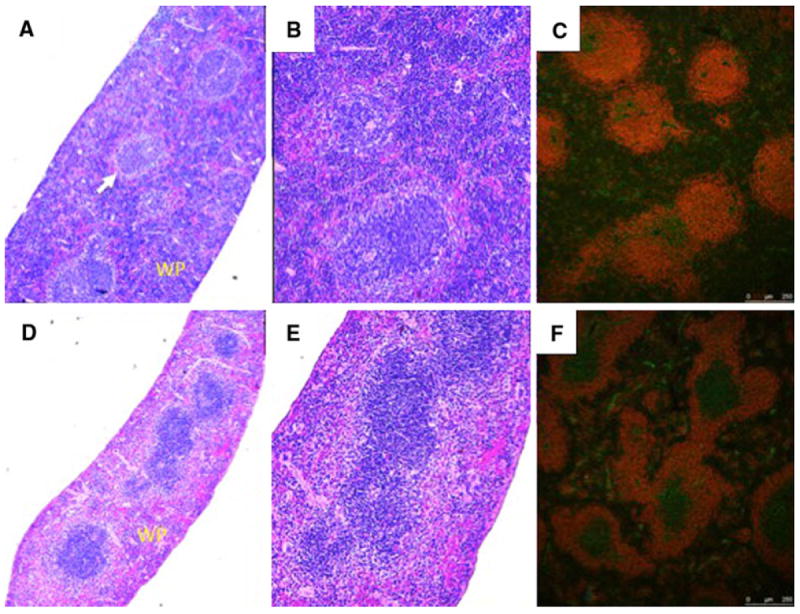
Fig. 3.

Histology and immunofluorescence staining of Ednrb+/+ and Ednrb−/− spleens. Ednrb+/+ a, b, c (upper panel) and Ednrb−/− d, e, f (lower panel). H and E staining (a, b and d, e); a and d (40×), b and e (100×) show significantly reduced white pulp (WP) with lymphopenia, fewer primary follicles and poorly defined marginal zone (arrow) in the Ednrb−/− (d and e) compared to Ednrb+/+ (a and b). Immunofluorescence staining for CD45R and CD3 of Ednrb+/+ (c) and Ednrb−/− mice (f). CD45R labels B cells red; CD3 labels T cells green
