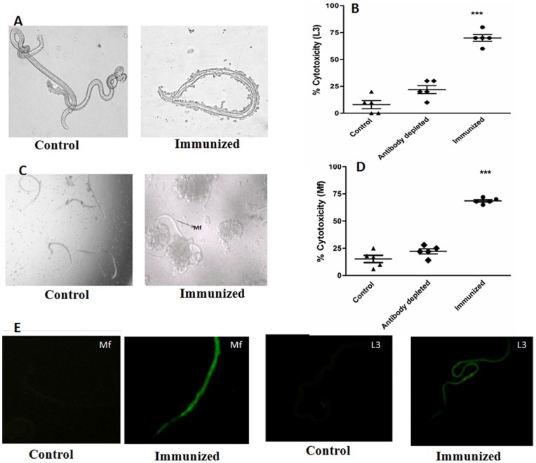
Figure 2. Antibody dependent cellular adhesion to L3 and Mf of B. malayi.
Mf and L3 were incubated with peritoneal exudates cells in the presence of anti Bm-TPP sera. Significant cellular adhesion on the surface of (A) L3 and (C) Mf was observed which caused death of parasite with in 48 h as a result cytotoxicity (B) L3 and (D) Mf. Photographs were captured on a phase contrast microscope (Nikon, Japan). Data are presented as mean ± S.E. of six replicate of experiment. Statistical significance based on the differences between the mean values among the groups are indicated as *p<0.05; **p<0.01 and ***p<0.001. (E) Interaction of anti Bm-TPP antibodies with B. malayi infective larvae (L3), and microfilariae (Mf) is demonstrated by indirect fluorescence. Parasites were incubated with anti Bm-TPP sera and further incubated with FITC labelled anti-mouse IgG. Images were captured under a fluorescent microscope at 20X for Mf, 10X for L3. Serum from naive animals at similar dilution was used as control and no detectable fluorescence could be seen in any of the above parasite stages.

