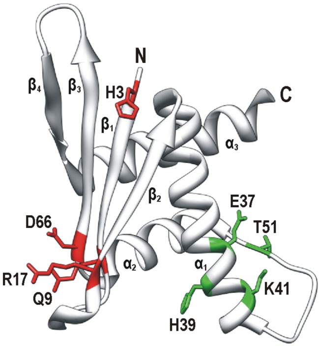
Figure 5. Amino acid residues of human ERH critical for its recruitment to nuclear speckles and replication foci.

Three-dimensional structure of a monomer of ERH was produced by UCSF Chimera package using coordinates from Protein Data Bank (2nmlA) [30]. Four β strands (β1, β2, β3 and β4), three α helices (α1, α2 and α3) and the N- and C-termini are indicated. Critical residues are shown with their side chains in color and are labeled using a single-letter code and position number in the polypeptide chain. Residues involved in the recruitment to nuclear speckles (in red) and replication foci (in green) are situated on the β sheet and near the loop between helices α1 and α2, respectively.
