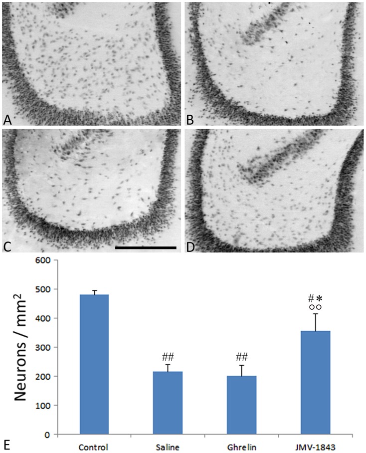
Figure 5. Photomicrographs illustrating neuronal cell loss in the hilus of dentate gyrus after status epilepticus (SE), in pilocarpine-treated rats.
Neurons were identified by the anti-neuron-specific nuclear protein (NeuN) antibody, as shown in the control staining (A). NeuN-immunopositive cells were markedly decreased in pilocarpine-treated rat of the saline- (B) and ghrelin-treated groups (C), sacrificed 4 days after SE. This phenomenon was significantly counteracted by JMV-1843 administration (D). Neuronal cell counts are shown in E. # = P<0.05, ## = P<0.01 vs the control non-epileptic group, * = P<0.05 vs the saline group, °° = P<0.01 vs the ghrelin group, Fisher's LSD test. Scale bar, 300 µm.

