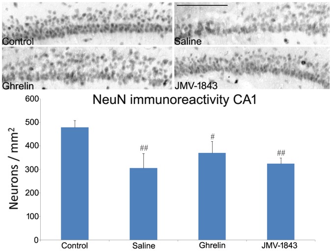
Figure 7. Photomicrographs illustrating neuronal cell loss in the CA1 hippocampal region after status epilepticus (SE), in pilocarpine-treated rats.
Neurons were identified by the anti-neuron-specific nuclear protein (NeuN) antibody, as shown in the control staining. NeuN-immunopositive cells were markedly decreased in pilocarpine-treated rat of the saline-treated group, sacrificed 4 days after SE. This phenomenon was not attenuated by treatment with GH secretagogues. # = P<0.05, ## = P<0.01 vs the control non-epileptic group, Fisher's LSD test. Scale bar, 150 µm.

