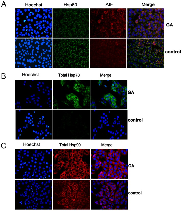
Figure 4. Effect of GA on localization and protein level of HSPs in OS 143B cells.
OS 143B cells were treated with 4 µM GA for 6 h; the levels of Hsp60, Hsp70, total cellular Hsp90 protein were determined by immunofluorescence. A. GA did not change the mitochondrial localization of Hsp60, however, it decreased the mitochondrial pool of the protein. Cell nuclei were shown in blue while Hsp60, AIF immunoreactivities in green and red, respectively. Merged image of all kinds of staining (orange) was also presented. B. GA upregulated Hsp70 protein level. Cell nuclei were shown in blue, while Hsp70 immunoreactivity in green. Merged image of both was also presented. C. GA upregulated Hsp90 protein level. Cell nuclei were shown in blue while Hsp90 immunoreactivity in red. Merged image of both was also presented. Original magnification ×40. Each experiment was performed at least three times. The representative data were shown.

