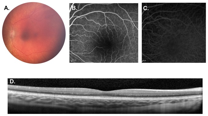
Figure 1. Weill Cornell LINCL Ophthalmic Severity Score 1.
A. Dilated fundus photograph of patient 14 showing normal appearing optic nerve, vessels and fovea. B. Mid-phase FA and C. ICGA of patient 20 appear normal. D. SD-OCT of the same patient demonstrates normal retinal architecture without disruption of the outer retina. FA – fluorescein angiogram, ICGA - indocyanine green angiogram, SD-OCT – spectral domain optical coherence tomography.

