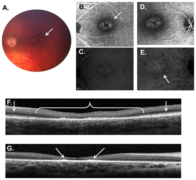
Figure 4. Weill Cornell LINCL Ophthalmic Severity Score 4.
A. Dilated fundus photograph of patient 9 shows a “bull’s eye” maculopathy with concentric rings of pigmentary changes emanating from the fovea (arrow). Some optic nerve pallor with attenuation of retinal vessels is also noted. B. Late phase FA of the right eye of patient 9 showing the “bull’s eye” maculopathy. C. Late phase FA of the left eye of patient 8 demonstrates a similar pattern within the macula. D. Late phase ICGA of the right eye of patient 9 correlates with the FA. E. Late phase ICGA of the left eye of patient 8. F. SD-OCT of patient 8 demonstrates considerable outer retinal abnormalities (*), including outer retinal atrophy and buildup of hyper-reflective material at the level of the retinal pigment epithelium. This outer retinal disruption typically extends less than 2 disc diameters (*), with normal appearing retinal architecture beyond the central fovea (arrows). G. Interestingly, the hyper-reflective material in some patients (including patient 16 shown here), appears to preferentially accumulate in the immediate parafoveal region (arrows). The late-phase FA (B and D) and ICGA (C and E) images show the hyper- and hypo-fluorescence areas (arrows) corresponding to the pigmentary changes on the color photograph (A) and the outer retinal abnormalities noted on the SD-OCT (F and G). FA – fluorescein angiogram, ICGA - indocyanine green angiogram, SD-OCT – spectral domain optical coherence tomography.

