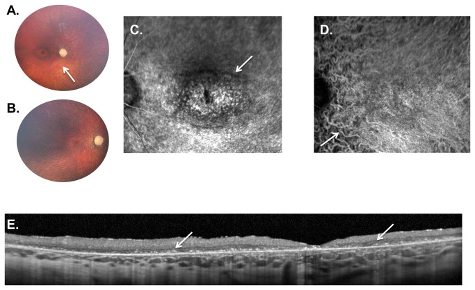
Figure 5. Weill Cornell LINCL Ophthalmic Severity Score 5.
A. Dilated fundus photograph of patient 4 shows a “bull’s eye” maculopathy with concentric rings of pigmentary changes emanating from the fovea. The optic nerve is completely pale with severe attenuation of retinal vessels (arrow). B. In patient 19, with most severe neurological score in this series, the retina is completely atrophied with a pale optic nerve and barely recognizable retinal vessels. A “bull’s eye” maculopathy is not evident on the color photography in this severely affected patient. C. Late-phase FA and D. ICGA show a large “bull’s eye” maculopathy with concentric rings of hypo- and hyperfluorescence (arrow). Due to the retinal atrophy, the choroidal vessels are prominent throughout the ICGA (D, arrow). E. SD-OCT of patient 19 demonstrates considerable outer retinal atrophy with buildup of hyper-reflective material at the level of the retinal pigment epithelium. This outer retinal disruption extends throughout the entire fundus without normal appearing retinal architecture anywhere in the fovea. FA – fluorescein angiogram, ICGA - indocyanine green angiogram, SD-OCT – spectral domain optical coherence tomography.

