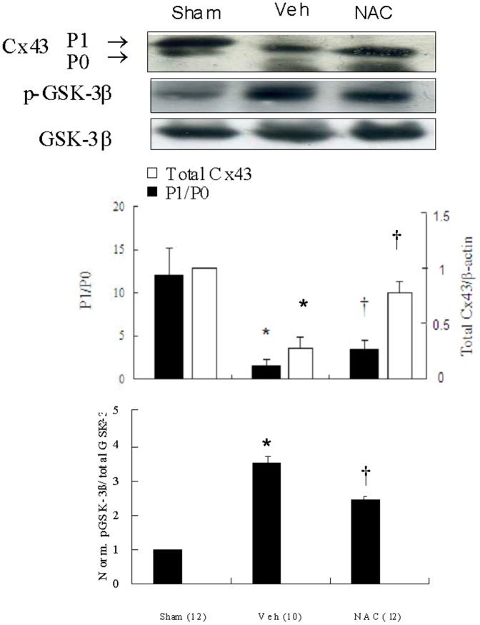
Figure 2. Western blot analysis of Cx43 and GSK-3β.
Ventricular remodeling after MI was associated with marked decreased amount of phosphorylated Cx43 (P1). Significantly increased Cx43 amount and P1/P0 ratio had taken place in the NAC-treated group compared with vehicle (Veh). However, a significantly decreased p-GSK-3β is noted in the NAC-treated group compared with vehicle. Relative abundance was obtained by normalizing the density of Cx43 protein against that of β-actin. Densitometric quantification of phosphorylation levels was expressed as the ratio of the density of phosphorylated band over total GSK-3β. Each point is an average of 3 separate experiments. P0, nonphosphorylated Cx43; P1, phosphorylated Cx43. *p<0.05 compared with sham and NAC-treated group; †p<0.05 compared with sham.

