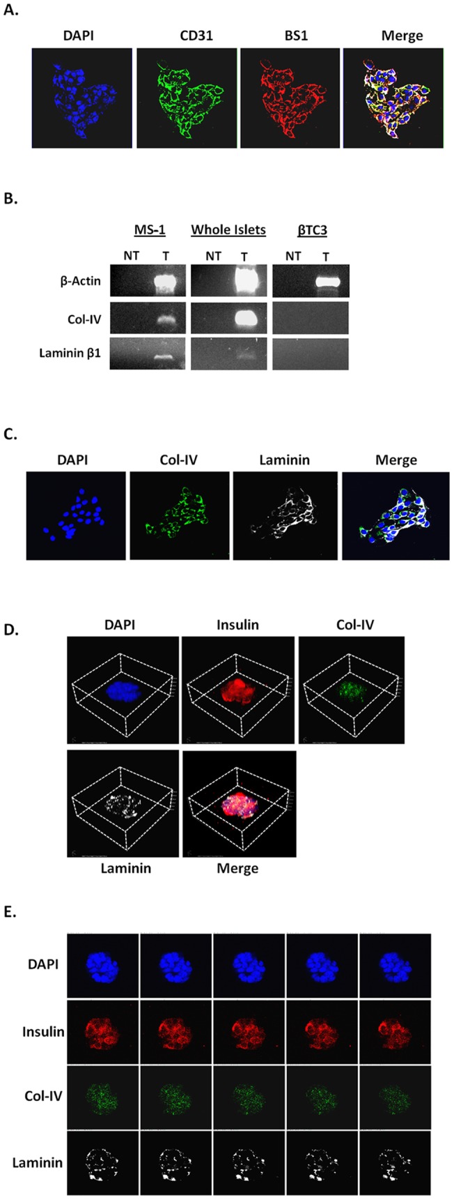
Figure 3. Col-IV and laminin are detected in and around the PI.

A. IF staining of MS1 cells. Blue-DAPI, Green-CD31, Red-BS1 and merge. White/Yellow represents double positive cells. B. RT-PCR for laminin β1 and col-IV in MS1, whole murine islet preps, and βTC3 cells. Laminin α1 and α2 were not detected (data not shown). C. IF staining of MS1 cells. Blue- DAPI, Green- col-IV, White-Laminin, and merge. D. 3-D reconstruction of z-stack imaging of an 8 d old PI. E. Non-consecutive z-stack confocal images of a PI. Blue- DAPI, Red- Insulin, Green- col-IV, White- laminin.
