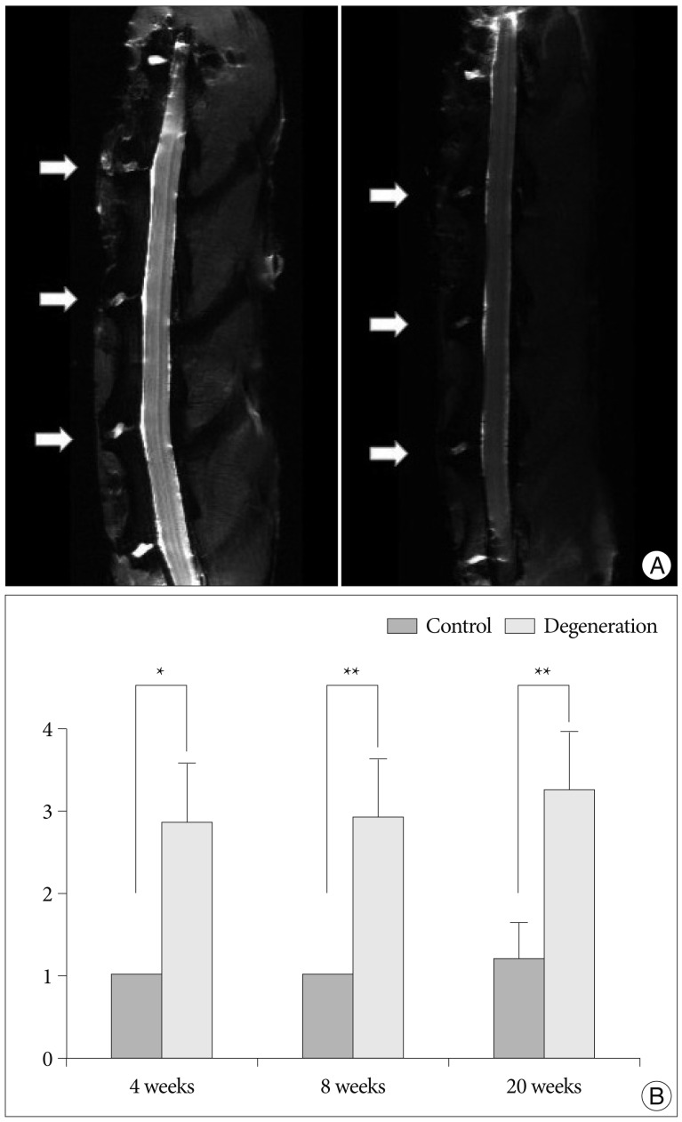
Fig. 4.
Ex vivo analyses of MRI of the rabbit spine. A : Representative T2-weighted sagittal image of lumbar spine shows that moderate degenerated discs (arrows; left) at L2-3, L3-4, and L4-5. Severe degenerated disc demonstrates more decreased in signal intensity and area (arrows; right). B : The MRI grade of the punctured discs is significantly increased compared to that of the control disc (L5-6) at each time period (*p<0.01, **p<0.001).

