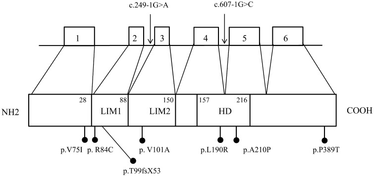
Fig. 1.
Schema of LHX4 genomic organization and protein structure. The location of reported mutations is shown. Mutations of introns are indicated by the arrows. Missense mutations and a frameshift mutation are indicated by black dots. Numbers next to the protein indicate amino acid positions of domain boundaries. White boxes, exon; LIM, LIM domains; HD homeodomain.

