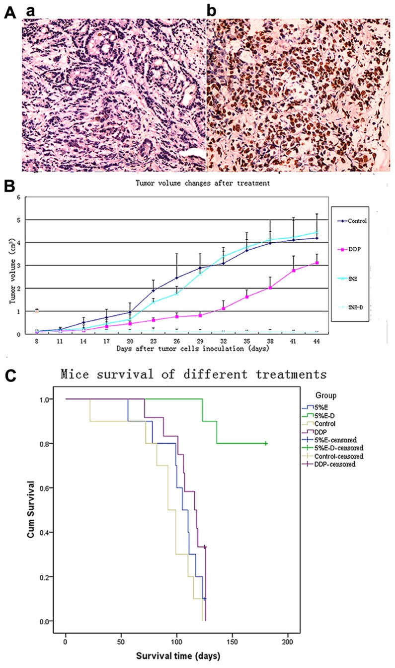
FIGURE 3.
(A) Apoptosis analysis of 5% ethanol–DDP-treated tumor tissues with TUNEL staining. (a) Control tumor tissue. (b) 5% ethanol–DDP treated tumor tissue. Compared with control, 5% ethanol–DDP significantlyIn artworks of Figure 3 has no part whereas the parts (A) and (B) explained in the caption. Kindly advise. increased tumor cell apoptosis rate (60.11% ± 7.52% vs. 5.32% ± 1.76%, p < 0.05). (B) DDP-resistant tumor size changes by various treatments.5% ethanol–DDP significantly inhibited tumor growth compared with control or DDP alone or 5% ethanol after 4 weeks’ treatment. E–D stands for ethanol–cisplatin. (C) Survival of A549/DDP tumor-bearing mice in different groups. 5% ethanol–DDP treatment significantly improved estimated mean survival time compared with control or DDP alone or 5% ethanol. E–D stands for ethanol–cisplatin.

