
Figure 4. Trichrome-Stained Transmural Section From the Left Ventricular Free Wall in a Control Dog and a Dog Treated With Alginate Hydrogel Implant.
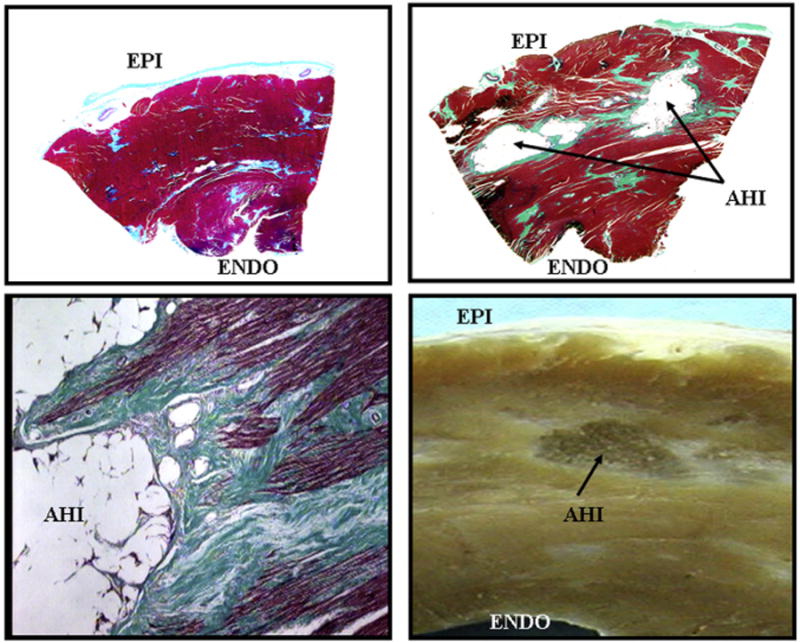
Trichrome-stained transmural section from left ventricular free wall in a control dog (upper left) and a dog treated with alginate hydrogel implant (AHI) (upper right) at end of study. AHI are encapsulated in thin layer of connective tissue (light green). Bottom left: high magnification trichrome-stained myocardial section showing the border between myocardium and AHI. Bottom right: photograph of gross LV free wall formalin-fixed section showing an AHI within the wall. EPI = epicardial surface; ENDO = endocardial surface; other abbreviations as in Figure 2.