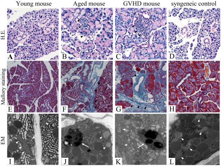
Figure 1. Light microscopic findings and electron microscopic findings of lacrimal glands of aged and cGVHD model mice.
Light microscopic findings of the lacrimal gland (A–H). Hematoxylin and Eosin staining of the young mice (A), aged mice (B), cGVHD mice (C), and syngeneic control mice (D). Note that mononuclear cells infiltration is found in the periductal areas of the aged (B, arrowheads) and cGVHD mice (C, arrowheads), whereas only a small number of mononuclear cells are found in the young (A) and syngeneic control mice (D). Mallory staining of the lacrimal gland (E–H). Note that excessive fibrosis (arrowheads) and mononuclear cells infiltration are found in the periductal areas of the aged (F) and cGVHD mice (G) but absent in the young (E) and syngeneic control mice (H). Electron microscopic findings of the lacrimal gland (I–L). Several mitochondria in the aged (J, arrowheads) and cGVHD mice (K, arrowheads) show swollen morphology and disorganization of cristae, whereas those in the young or syngeneic control mice do not show any phenotypic alterations (I, L, arrowheads). Lipofuscin formation is found in the aged (J, arrow) and cGVHD mice (K, arrow) but absent in the young and syngeneic control mice (I, L). H.E.; Hematoxylin and Eosin staining, EM; Electron microscopy. Original magnification. A, D, E, X100, B, C, F–H, X200, I–L, X15000.

