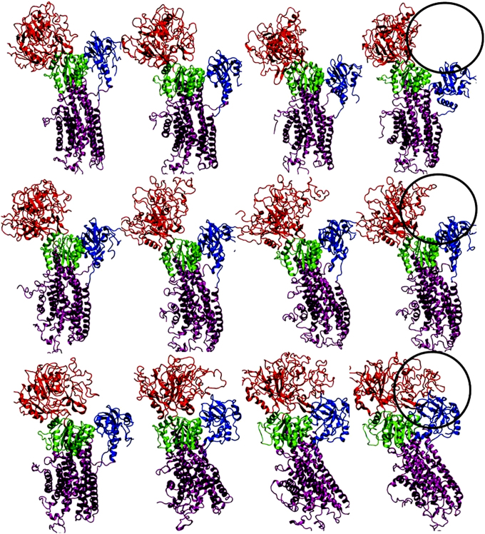
Figure 2. MD snapshots generated from individual trajectories after every 25 ns for PfATP6 enzyme (top row), artemisinin bound PfATP6 (second row) and Fe-artemisinin adduct bound PfATP6 (third row).
Nucleotide binding (N) domain and the actuator (A) domains are seen to be closing in the case of Fe-artemisinin adduct bound PfATP6 system.

