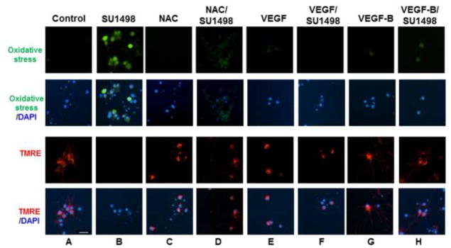
Figure 4. VEGF and VEGF-B protect against oxidative stress and mitochondrial dysfunction induced by SU1498.

Neurons were cultured in NB for 48 hr in the absence and presence of NAC (5 mM, final 24 hr), VEGF or VEGF-B (100 ng/ml for 48 hr, replenished after 24 hr). After 46 hr of incubation, cells were treated with SU1498 as indicated and labeled with either carboxy-H2DCFCA to detect oxidative stress (green) or TMRE (red) to measure the Δψm as described in “Materials and methods”. Nuclei were counterstained with DAPI. Scale bar: 50 μm. Data are representative of experiments that were repeated at least three times.
