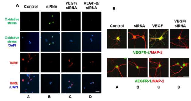
Figure 5. VEGF and VEGF-B protect against oxidative stress and mitochondrial dysfunction induced by siRNA VEGFR-2.

(A), Neurons were transfected with siVEGFR-2 as described in “Materials and methods” and then cultured without and with VEGF or VEGF-B for oxidative stress and TMRE measurements as described in Figure 4. Scale bar: 50 μm. (B), Cells were double labeled for fluorescence with MAP-2 (red) and either VEGFR-2 (green; upper panels) or VEGFR-1 (green; lower panels) as described in “Materials and methods”. Scale bar: 100 μm. Data are representative of experiments that were repeated at least three times.
