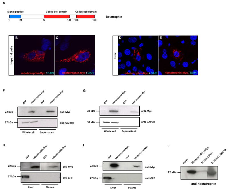
Figure 4. Betatrophin encodes a secreted protein.
(A) Predicted domains of the betatrophin protein. Cellular localization of mbetatrophin-Myc (B) or hbetatrophin-Myc protein (C) when transfected into the liver cell line Hepa1-6. Cellular localization of mbetatrophin-Myc (D) or hbetatrophin-Myc (E) when overexpressed in mouse liver through hydrodynamic tail vein injection. Western blots show mbetatrophin-Myc protein (F) or hbetatrophin-Myc (G) in the supernatant following gene transfection into 293T cells. GFP gene transfection and the intracellular GAPDH protein are used as controls. Western blots show mbetatrophin-Myc (H) or hbetatrophin-Myc (I) protein in plasma (3 days after injection) when the gene is overexpressed in mouse liver by hydrodynamic tail vein injection. GFP gene injection is the negative control. (J) Western blot of human betatrophin in human liver and plasma samples. Cell lysate of hbetatrophin-Myc transfected 293T cells is the positive control and cell lysate of GFP transfected 293T cells is the negative control.

