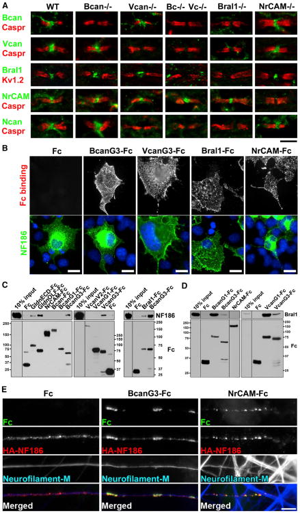
Figure 2. Node-Enriched ECM Components Interact with and Cluster NF186.
(A) Adult WT and mutant mouse spinal cord immunostained using antibodies against various ECM components (green) and the paranodal marker Caspr (red) or the juxtaparanodal marker Kv1.2 (red). Scale bar, 10 mm for Bral1 staining and5 mm for all the others.
(B) Fc fusion proteins of BcanG3, VcanG3, NrCAM, and Bral1 bind to COS-7 cells transfected withHA-NF186 (green). Fc alone was used as a negative control. Cell nuclei were visualized by Hoechstin blue. Scale bars = 20 μm.
(C) Pull-down analysis shows binding between BcanG3, VcanG3, or Bral1 and the secreted NF186 extracellular domain. GldnECD is the whole extracellular domain of gliomedin, and GldnOLF is the olfactomedin domain of gliomedin; both serve as positive controls. Molecular weight markers in kDa.
(D) Pull-down analysis shows binding between BcanG1 or VcanG1 and Bral1.
(E) Clustering of BcanG3-Fc or NrCAM-Fc (green) and HA-NF186 (red) along axons (visualized by anti-neurofilament-M) of cultured DRG neurons. Scale bar, 5 μm.
See also Figure S1 and Table S1.

