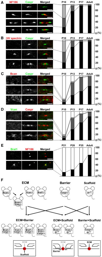
Figure 3. Developmental Clustering of Nodal and Paranodal Components in the CNS.
(A–E) Rat optic nerve sections were immunostained with antibodies to nodal or paranodal markers as indicated in the left panels showing representative images of different stages of CNS node formation. Scale bars, 5 μm. Right panels show a quantitative analysis of each type of staining as a function of age. The data were obtained by observation of 200–250 sites from two animals at each time point indicated.
(F) Schematic of the genetic strategy used to test whether multiple mechanisms contribute to CNS node of Ranvier formation. ECM mutants, PJ mutants (barrier), and CS mutants (scaffold) were crossed to generate double- or triple-mutant mice with two mechanisms disrupted simultaneously. (N.B. The core ECM molecules at the CNS nodes cannot be completely removed in ECM+Barrier or ECM+Scaffold mutant mice.)

