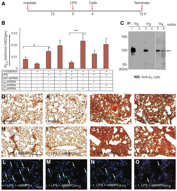
Figure 4.
Silencing of α4 or α5 integrin abolishes mBMPCs-mediated restoration of lung fluid balance. (A): Timeline used to determine capillary filtration coefficient (Kfc) following treatment with mBMPCs. (B): Kfc assay following treatment with mBMPCs, mBMPCs-α5-shRNA, or mBMPCs-α2-shRNA or mBMPCs-α4-shRNA (n = 10 per group). Experiments were performed five times. *, p < .05. **, p < .01. (C): shRNA-mediated silencing of α2, α4, and α5 integrins was determined by immunoprecipitation followed by WB with indicated antibodies. (D–H): H&E-stained lung sections: (D) normal alveolar septa with no obvious PMN infiltration; (E) normal alveolar septa with no obvious polymorphonuclear (PMN) infiltration in response to γ-irradiation; (F) septal thickening indicative of edema in response to γ-irradiation and LPS treatment; (G) no protection with mECs, uptake of tissue PMN, and edema formation; (H) normal alveolar septa with no PMN infiltration in mouse lung following mBMPC treatment; (I) silencing of α2 integrin fails to abolish effect of mBMPCs, as illustrated by normal lung architecture; and (J, K) silencing of either α4 or α5 integrin offered no therapeutic benefit to mice that were previously injured by irradiation and LPS. (L–O): Retention of mBMPCs expressing RFP was examined at the end of 48 hours by staining with an anti-RFP polyclonal antibody (green). White arrows indicate mBMPCs expressing RFP. For quantification, please see supporting information Figure S7. Data are representative of at least three independent experiments. Magnification 200×. Scale bar 150 μm. Abbreviations: ECs, endothelial cells; LPS, lipopolysaccharide; mBMPCs, mouse bone-marrow-derived progenitor cells; RFP, red fluorescent protein; WB, Western blotting.

