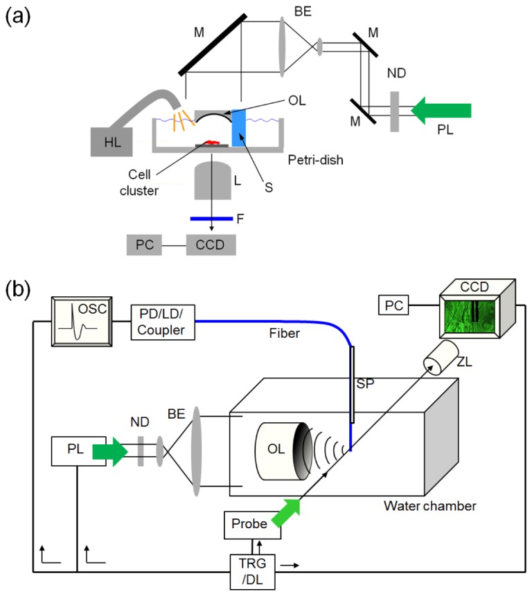
Fig. 1.

Experimental schematics. (a) A setup for micro-fractionation of cell clusters by LGFU. The setup was prepared on the inverted microscope (BE: beam expander, F: optical filter, HL: halogen lamp, L: objective lens, M: mirror, ND: neutral density filter, OL: optoacoustic lens, PL: Nd:YAG pulsed laser beam (6-ns pulse width), S: supporting frame,). (b) A shadowgraphic imaging setup (LD: laser diode, OSC: digital oscilloscope, PD: photodetector, Probe: probe laser beam (1-ns pulse width), SP: supporting plate, TRG/DL: trigger and delay-generator unit, ZL: zoom lens).
