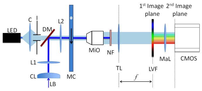
Fig. 1.

Experimental setup. A laser beam (LB) induces fluorescence in a cell flowing through the channel (MC) and is then blocked by the notch filter (NF). Fluorescence is filtered by the LVF and is then imaged onto the camera (CMOS) through a macro photography lens (MaL). The LED light, collimated by a condenser and an iris (C and I), is transmitted by a portion of the LVF and provides a bright field image of the cell. L1 and L2 form a telescopic system and MiO is a microscope objective followed by the tube lens TL.
