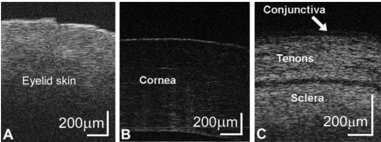
Fig.6.

Real-time images of (a) eyelid skin, (b) cornea, and (c) conjunctiva. (b) The outer corneal epithelium and inner Descemets membrane are visible in the corneal images. (c) The layers of conjunctiva and Tenons are observed above the dense sclera.
