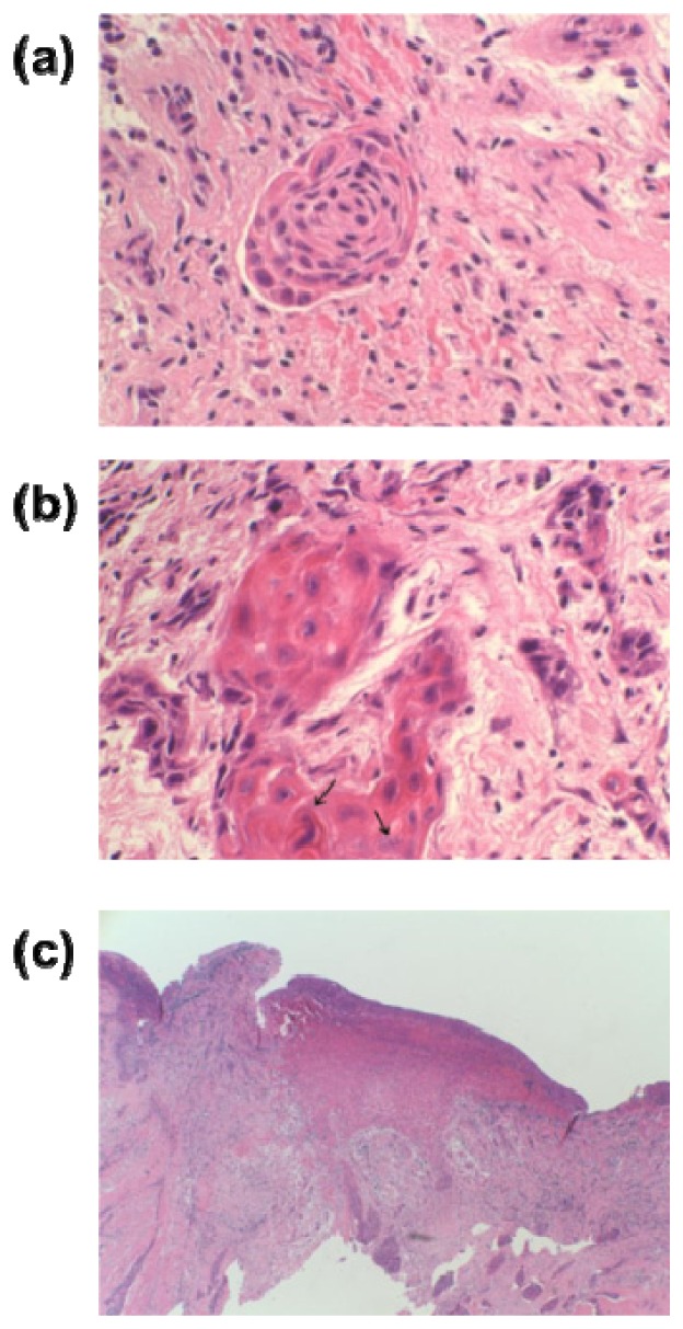
Fig. 5.

(a) A nest of deformed mucosal cells with hyperchromatic nuclei from sample #1 ( × 400), (b) a mixture of inflammatory cells from sample #2 ( × 400), (c) mucoepidermoid carcinoma of the tongue showing subepithelial invasion from sample #1 ( × 40).
