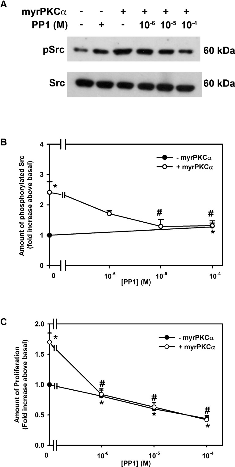
Figure 10.
Effect of inhibition of Src on constitutively active PKCα-stimulated cultured rat conjunctival goblet cell Src phosphorylation and proliferation. Cultured rat conjunctival goblet cells were serum starved for 24 hours and preincubated with PP1 (10−6–10−4 M) for 30 minutes prior to addition of an adenovirus containing myristolyated (active) PKCα (myrPKCα) (1 × 10⁷ pfu) or with no addition for 22 hours. The amount of total and phosphorylated Src was determined by Western blot analysis, and a representative blot of three independent experiments is shown in (A). The blots were scanned, and the data shown in (B) represent mean ± SEM of three experiments. The number of proliferating cells was determined by WST-8 and is shown in (C). Data are mean ± SEM from three independent experiments. *Statistically significant difference from basal, set to 1.0. #Statistically significant difference from myrPKCα.

