
Figure 4.
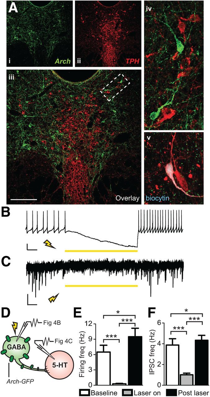
Archaerhodopsin-mediated photoinhibition of DRN GABAergic neurons silenced their spontaneous firing and decreased IPSC activity of neighboring 5-HT neurons. Ai–Aiv, Confocal image of Arch-GFP-expressing GABAergic neurons (green) and TPH labeled 5-HT neurons (red) in the DRN. Scale bar, 50 μm. Av, Example of a 5-HT neuron recorded in the DRN that was filled with biocytin and its neurochemical identity was confirmed a posteriori using TPH immunostaining. B, Current-clamp trace of a spontaneously firing, Arch-expressing GAD2+ GABA neuron. Exposure to 318 mW mm−2 of 593 nm light inhibited the tonic firing. Scale bar, 20 mV, 1 s. C, Voltage-clamp recording from a later identified 5-HT neuron. Illumination of the DRN with 318mW mm−2 of 593 nm light resulted in a reduction of IPSC frequency that returned upon cessation of laser stimulation. Scale bar, 10 pA, 1 s. D, Schematic of recording experiments. Whole-cell patch-clamp was performed on either GABA or 5-HT neurons while Arch-expressing GABA neurons were photoinhibited via laser stimulation. E, Summary histogram showing that laser illumination resulted in complete silencing of spontaneous firing of GAD2+ GABA neurons [N(n) = 3(18)] that was restored upon laser cessation (repeated-measures ANOVA, Fisher's post hoc; baseline vs laser, ***p < 0.001; laser vs post-stim ***p < 0.001; baseline vs post-stim *p = 0.030). F, Summary histogram showing that laser stimulation reduced IPSC frequency in 5-HT neurons [N(n) = 2(5)] that was restored after laser cessation (repeated-measures ANOVA, Fisher's post hoc; baseline vs laser ***p < 0.001; laser vs post-stim ***p < 0.001; baseline vs post-stim *p = 0.047).